Saturday, February 23, 2013

Carbohydrates

Sugar structure 
MONOSACCHARIDE 
Glucose
Glucose (or Glucopyranose) is the most common sugars which has two different form, alpha and beta.
α-D-Glucose and β-D-Glucose
Galactose
Galactose is similar to Glucose, however, the fourth hydroxide on the ring projects upward
α-D-Galactose and β-D-Galactose
Fructose
A furanose ring structure with six carbons or (hexose)
α-D-Fructose and β-D-Fructose
Ribose
A furanose ring structure with five carbons or (pentose) 
α-D-Ribose and β-D-Ribose
N-acetylglucosamine (NAG)
It is a glucose with a "peptide-like" bond attaches on the second carbon
N-acetylglucosamine
β-D-Glucuronate 
β-D-Glucuronate
DISACCHARIDE 
Amylose (2 and more α-glucose)
It is a disaccharide which is created by α 1->4 glycosidic linkage.
The structure of amylose is a left handed helix, 6 glucoses/ turn, and -CH2OH groups project outward 

Two different projections of Amylose
Cellobiose  (2  β-glucose) 
It is created by β 1-> 4 glycosidic linkage. With more than 2 glucose, this chain will form Cellulose 


Chair Conformation of Cellobiose
Source: http://nl.wikipedia.org/wiki/Bestand:Cellobiose_skeletal.png

Cellobiose 
β-D-Lactose (β-Galactose and β-Glucose)
A sugar that found in milk, which is created by a β 1->4 glycosidic linkage 
Lactose molecules
Source: http://www.edinformatics.com/math_science/science_of_cooking/lactose.htm
Sucrose (α-Glucose and β-Fructose) 
Notice that the fructose molecule need to rotate to the left 180 degrees, around the z-axis (the axis that projects out of the plane). It is a non reducing sugar. 
Sucrose
Source: http://users.bergen.org/dondew/bio/AnP/Anp1/AnP1Tri1/CARB_ART/SUCROSE_SYNTH/SucroseSynth.html
Trehalose (α-Glucose and α-Glucose)
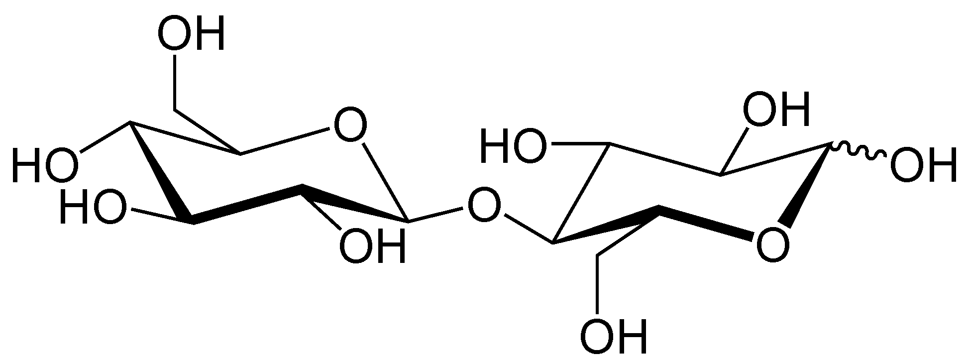
 It is animal blood sugar which is linked by α 1->1  glycosidic linkage 
Note: The right handed side glucose must rotate 180 degree to the left around the z-axis (the axis that projects out of the plane). 
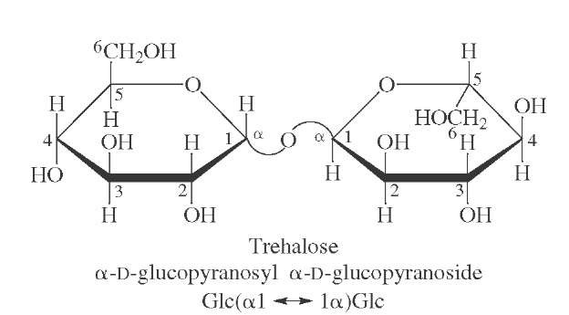
Trehalose Haworth projection
Source: http://what-when-how.com/glycoconjugates-and-carbohydrates/polysaccharides-glycoconjugates-and-carbohydrates/
Source: Dr. Larry Jon Friesen' s Lectures

Saturday, February 16, 2013

Enzymes

Classification of Enzymes 
1) Oxidoreductase
2) Transferase
3) Hydrolase 
4) Lyase
5) Isomerase
6) Ligase

Ex: Hexokinase 2.7.1.1
A Stereotypical diagram of the Holoenzymes
Inhibition of Enzymes 
There are three different Inhibitions, Competitive inhibition, Noncompetitive inhibition, and uncompetitive inhibition.


Competitive Inhibition: 
The Inhibitor binds to the catalytic center or active site of the enzyme and prevents the substrate from binding on the enzymes. Eventually, the Michealis constant (Km) will be higher than the stage without any inhibitors. 
Diagram of Competitive Inhibitor


Figure 1 Competitive Inhibition
According to Michealis, it is hard to determine the Vmax by looking at the hyperbolic graph. Therefore, he took the reciprocal of the concentration of substrate and the velocity, which allowed him to graph the inverse of hyperbola or Linear. As shown in the Figure 1, the Vmax of the process can be determined by the y-intercept of the graph. 

NOTE: Competitive Inhibition: Vmax the same, Km Increase.


Source: Dr. Larry Jon Friesen' s Lectures 

Useful Link: http://www.rcsb.org/pdb/home/home.do

Proteins

*Protein Structures 
Primary Structure 
All amino acids are connected by peptide bond (O=C=N-H)
Due to the high electronegativity of Nitrogen and Oxygen, the electrons are pulled from the carbon atom toward them which form a resonance structure (C=N) and (C=O). The possibility to form C=O bond is 60% and 40% for C=N
Characteristics of peptide bond: Polar, Resonance, Non-rotatable, Planar
Primary Structure of a pentapeptide in NCC sequence
Primary structure can be written in NCC sequence or CCN sequence, which represent the same structure. Note: the orientation is very important
In NCC sequence: the first Hydrogen on alpha-carbon projects in the planar then alternating (in and out)

Secondary Structure
All primary structures are connected by hydrogen bond

Alpha-Helix characteristics  
Alpha-Helix Structure
Source: Dr. Larry Jon Freisen's Lab Manual 
1)Right handed helix
2)R-groups project outward
3)Rise angle 27 degrees
4)3.6 amino acids per turn
5)0.15nm rise
6)0.51nm interchain distance
7)0.54nm pitch
8)"Strechy"
9)Angle subtented by amino acids= 100 degrees
10)Hydrogen bonding between peptide bonds

Beta-Pleated Sheet
The folded shape of Beta-Pleated sheet allows the R groups project up and down which will able to form the tertiary and quaternary structure with the R-Group Interactions

Beta-Pleated sheet with R groups project up and downward 

Tertiary Structure 
All secondary structures are connected by the R-Group Interactions

Quaternary Structure 
All tertiary structures are connected by the R-Group Interactions


*R-Group Interactions 
Non-Covalent Type 
1) Hydrogen Bonding
Serine interacts with Glutamine under Hydrogen bonding

2) Dipole-Dipole Bonding
Serine interacts with Serine under Dipole moment 
3) Ionic Bonding
The cation of one amino acid interacts with the anion of other amino acid
Ammonium ion of Lysine interacts with acetate ions on Aspartate 

4) Cation-Pi Bonding 
The resonance of Pi bond has created electron clouds, which are able to attract the Positive cations from other amino acids.
The Electron Cloud of Phenylalanine attracts the cation of Lysine

5) Hydrophobic Bonding 
Phenylalanine is nonpolar molecule which is hydrophobic
Under the surface of tension water sphere, the electrons in electron clouds have been pushed up to the upper clouds, which created a relative strong bond.
Phenylalanines bond together under water tension
Covalent Type
1) Isopeptide Bonding 
This kind of bonding is similar with peptide bonding; however, it is not on the main chain but on the between the amine group and carboxylic group within the  R-groups. 

2) Disulfide Bonding (Only on 2 Cysteines) 
Two Cysteines joined together by releasing two protons and electrons.
Cysteine connects with Cysteine

Source: Dr. Larry Jon Friesen' s Lectures  

Sunday, February 3, 2013

Amino Acids

There are 20 essential amino acids in our body which can combine and form many different peptides and proteins. 
The structures of amino acids are determined by the pH of the solution. The pK1 (or pKa) is the point where caboxylic acid group (-COO-H) lose one proton (Hydrogen). At pK2 (pKb), the amine group (NH3+) will be deprotonated and form NH2. There are vary numbers of pKR, the point where the R-group will be deprotonated. 

Example: 
Aspartic Acid Titration Curve
Source: http://faculty.une.edu/com/courses/bionut/distbio/obj-512/Chap6-titration-aspartate.html